By Karly Cohen, for Friday Harbor Labs.
I have had the opportunity to study a diversity of structures and critters – from teeth and body armor like tough scales, to softer tissues like lips and the discs of fishes that suck onto surfaces. Each project requires a set of imaging tools that reveal hard and soft anatomy in different ways. One of the great joys of my work is translating careful observations of anatomy into beautiful images and figures. 2D visualization like histology and photography let me see the world through clearly arranged points of view, while 3D imaging shows me context and relationships.
I started as a histologist, someone who slices things thinly to allow stains to reveal different tissues. I use paraffin and plastic histology to measure qualitative and quantitative traits of tissues like how collagen is arranged or how much muscle is present to make predictions about their mechanics.
Pacus, the herbivorous cousins of notoriously carnivorous piranhas, have large multi-cuspid teeth that are hidden by large lips analogous to the tongues and snoots of giraffes and rhinos (Cohen and Kolmann, 2021). Using plastic histology, I sectioned the lips of the pacus to find that they are not muscular like those of mammals, but are instead composites with layers of collagen, fat and keratin (the material that makes up our fingernails). Sometimes it’s important to not just see the morphology and arrangement of tissues but also know their identity. This is where paraffin histology comes into play. It revealed that some of the soft tissue in pacu lips is elastin: a protein responsible for stretchiness.
Teeth and armor are inherently 3-dimensional structures, as they interact with food and the environment from many directions. 3D problems require 3D solutions and analyses; CT scanning lets us see reconstructed anatomy in high resolution without the artifacts created by dissection. The teeth I study are small. Really small. For instance, a single tooth of a bristlemouth fish — the most abundant vertebrate on the planet — is 30 microns thick, which is thinner than a strand of hair. CT scanning allows me to see these important feeding structures from a whole new point of view, and 3D print them 200% larger than they are in life. Doing this enabled me to show that not all small, pointy teeth work in the same way and that these seemingly simple dentitions are more complicated than the considerations of a single tooth. Curved teeth in the goosefish are used to puncture and hold onto prey, and the exaggerated teeth in the fangtooth fish effectively “cage” prey inside the mouth to prevent prey from swimming away (Cohen et al. 2020).
Scanning electron microscopy (SEM) lets you see the outside topography of structures and has been crucial in seeing damage on spines, scales and teeth. Damage is the scar left behind when critters interact with food or the environment, and it shows how a given structure is being used. The Pacific spiny lumpsucker is a favorite fish of FHL researchers, and gets its name from the conical armor that covers its whole body. With SEM we were able to see that the armor of lumpsuckers is often abraded with small fractures around the base. Abrasion wears away high points, leaving grooves and scratches that reveal the nature of the abrading surface. In contrast, impacts cleave or spall armor, leaving sharply defined edges and shatter patterns explained by the direction of the impact.
I have spent much of my time at FHL mastering different imaging tools, and having access to them has been instrumental in inspiring new ideas. My research would not be as broad or integrative if not for the opportunity to actually drive the machines responsible for these images. There are few research centers that provide access to both critters and the tools to visualize them, which is what makes FHL a truly special place.
Thank you to the Karel F. Liem Bioimaging Center at FHL and to the donors that made the establishment and upkeep of these instruments possible. Tools like these are costly, and often promote the idea that to do good and exciting science one must have extraordinary amounts of funding. At FHL, this equipment is free to use and available if you are willing to learn how to drive it, making our corner of campus a busy place 24/7/365. I am grateful for the critical funding I received from a Stephen and Ruth Wainwright Fellowship and from the FHL Marine Science Fund. This support allowed me to conduct research at FHL over the past 3 years and especially through a pandemic when much of my funding was unsure.
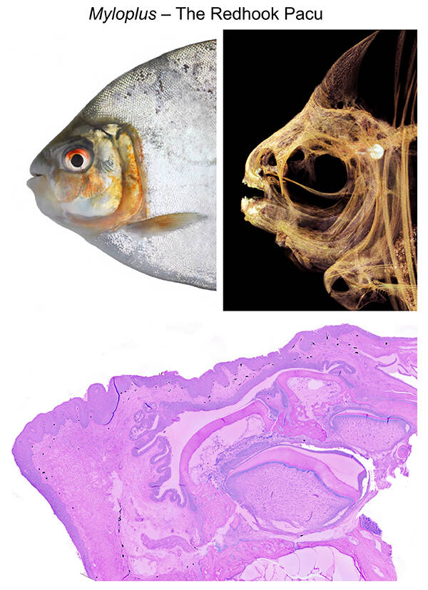
Contributed photo Left: The Redhook pacu, Myloplus, (adopted from Huie et al. 2019) vs. in CT. Below is a histology section through the lower lip revealing layers of collagen, keratin and other materials that help this species grab and hold onto food.
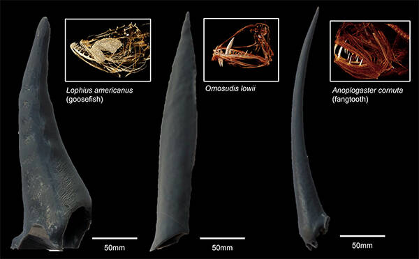
3D prints and CT scans of three different fishes with canine-like teeth. These 3D prints are scaled up from the original size of the fish, allowing us to interpret their shape with new perspective. (Contributed photo)
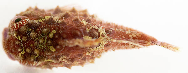
Top (dorsal) view of a Pacific spiny lumpsucker, a favorite fish at FHL and one of the many armored fishes we find in our local subtidal ecosystems. (Contributed photo)

Scanning electron microscope images of spiny lumpsucker scales. These images showcase damage that we find scarring the outside of lumpsucker armor. Looking at damage lets us make predictions about how the armor is used. (Contributed photo)